Featured Answer: How do I know if my dental implant is failing?
The most common warning signs of a failing dental implant include increasing pain or discomfort around the implant (especially after the initial healing period), swollen or bleeding gums around the implant site, an implant that feels loose or moves when you press on it, receding gum tissue that exposes the metal post, and persistent bad taste or odor near the implant. If you notice any of these symptoms, contact your dentist promptly, early intervention is the difference between saving the implant and losing it. At Innova Smiles in Marlborough, Dr. Fatima uses annual radiographs and probing measurements to detect bone loss around implants before symptoms appear.
Understanding Implant Failure: How Common Is It?
Dental implants are among the most predictable procedures in all of dentistry. According to the American Academy of Implant Dentistry (AAID), implants have a success rate exceeding 95 percent at 10 years. Long-term studies published in Clinical Oral Implants Research have tracked implants for 20 years and beyond, with cumulative survival rates of 90 to 95 percent at the 20-year mark.
But those statistics also mean that 2 to 5 percent of implants do fail, and when they do, knowing what to look for makes all the difference. An implant caught in the early stages of trouble can often be saved with non-surgical treatment. An implant that goes unmonitored until the bone around it has been destroyed may need to be removed entirely.
Implant failure falls into two distinct categories, early failure and late failure, and they have different causes, different timelines, and different warning signs.
Early Implant Failure (0-6 Months)
Early failure occurs before osseointegration is complete, that is, before the titanium implant has fully fused with the surrounding jawbone. This biological bonding process, first described by Professor Per-Ingvar Branemark in the 1960s, takes approximately 3 to 6 months. During this period, the implant is vulnerable because it has not yet formed a solid mechanical connection with the bone.
Early failure affects roughly 2 to 3 percent of implants, according to a 2019 systematic review in the Journal of Oral and Maxillofacial Research.
Causes of Early Failure
Failed osseointegration: The implant simply does not bond with the bone. This can happen when blood supply to the surgical site is inadequate, when the bone quality is poor (very soft or porous bone), or when the implant was placed with insufficient primary stability (the initial tightness of the implant in the bone at the time of surgery). A 2018 study in Clinical Oral Implants Research identified low insertion torque, a measure of how tightly the implant grips the bone at placement, as one of the strongest predictors of early failure.
Surgical site infection: Bacteria can contaminate the surgical site during or after implant placement, preventing the bone from healing around the implant. This is rare when proper sterile protocols are followed, but it can occur in patients with compromised immune systems, uncontrolled diabetes, or poor oral hygiene during the healing period.
Premature loading: If an implant is subjected to chewing forces before osseointegration is complete, the micro-movements can disrupt the bone-to-implant interface. This is why most implant protocols involve a healing period of 3 to 6 months before the final crown is placed. Immediate-load (same-day teeth) protocols exist but require specific bone conditions and careful case selection.
Overheating during surgery: Drilling into bone generates heat, and if the bone temperature exceeds 47 degrees Celsius for more than one minute, the bone cells can die (thermal necrosis), preventing osseointegration. This is avoided through proper drilling speed, copious saline irrigation, and sharp surgical drills, standard protocol at Innova Smiles.
Insufficient bone volume: If there is not enough bone height or width to fully encase the implant, the exposed portion cannot integrate. This is why Dr. Fatima uses CBCT 3D imaging to measure bone dimensions precisely before every implant case. When bone volume is insufficient, a bone graft or sinus lift is performed before or during implant placement to build the foundation.
Warning Signs of Early Failure
| Symptom | What It May Indicate | Urgency |
|---|---|---|
| Implant feels loose or mobile | Failed osseointegration | Call immediately |
| Increasing pain after the first week (pain should be decreasing) | Infection or bone necrosis | Call within 24 hours |
| Swelling that worsens after day 3-4 (should be improving) | Infection | Call within 24 hours |
| Pus or discharge from the surgical site | Active infection | Call immediately |
| Implant is visible through the gum or the gum has receded from the site | Implant migration or bone loss | Call within 48 hours |
| Numbness or tingling that persists beyond 48 hours | Possible nerve irritation | Call within 48 hours |
Patients from Hudson, Framingham, and Northborough who have had implants placed at Innova Smiles are scheduled for follow-up visits at 1 week, 4 weeks, and 3 months post-surgery. These visits allow Dr. Fatima to monitor healing, take periapical radiographs when indicated, and catch any signs of early failure before they progress.
Late Implant Failure (6 Months and Beyond)
Late failure occurs after the implant has successfully osseointegrated and been restored with a crown, bridge, or denture. The implant functioned normally for months or years before problems developed. Late failure is almost always caused by peri-implantitis (infection and bone loss around the implant), mechanical complications, or systemic health changes.
Peri-Implantitis: The Primary Cause of Late Failure
Peri-implantitis is a destructive inflammatory condition affecting the tissue surrounding an osseointegrated implant, resulting in loss of supporting bone. It is the implant equivalent of periodontitis (gum disease) around natural teeth, and it is the single most common cause of late implant failure.
How common is it? A 2018 meta-analysis published in the Journal of Clinical Periodontology, one of the most cited studies on the topic, estimated that peri-implantitis affects approximately 22 percent of implant patients and 10 percent of individual implants. These figures vary across studies depending on the diagnostic criteria used, but the consensus is that peri-implantitis is not rare.
What happens biologically: Bacteria colonize the implant surface at and below the gumline, triggering an inflammatory response. Unlike natural teeth, implants lack a periodontal ligament, the fibrous tissue that connects natural teeth to bone and provides a barrier against bacterial migration. This structural difference means that once bacteria penetrate beneath the gumline around an implant, the infection can progress to the bone more rapidly than around a natural tooth.
The bone around the implant begins to resorb (dissolve), creating deepening pockets that harbor more bacteria, which triggers more bone loss, a self-reinforcing cycle that, untreated, eventually destroys enough bone that the implant becomes loose and must be removed.
Stages of peri-implant disease:
-
Peri-implant mucositis, inflammation of the soft tissue around the implant without bone loss. This is the reversible stage, analogous to gingivitis around natural teeth. Symptoms include bleeding when probing, redness, and swelling. A 2017 consensus report from the World Workshop on the Classification of Periodontal and Peri-Implant Diseases confirmed that peri-implant mucositis is a precursor to peri-implantitis and that early treatment can prevent progression.
-
Peri-implantitis, inflammation with progressive bone loss. Once bone loss begins, the damage is not naturally reversible. Treatment aims to halt progression and, in some cases, regenerate lost bone through grafting.
Warning Signs of Late Failure
| Symptom | What It May Indicate | Urgency |
|---|---|---|
| Bleeding gums around the implant during brushing or probing | Peri-implant mucositis or peri-implantitis | Schedule appointment within 1-2 weeks |
| Red, swollen, or puffy gum tissue around the implant | Active inflammation | Schedule appointment within 1-2 weeks |
| Increasing pocket depth around the implant (measured by your dentist) | Bone loss in progress | Requires treatment plan |
| Bone loss visible on X-ray | Peri-implantitis confirmed | Requires treatment plan |
| Implant crown feels loose or rocks | Abutment screw loosening or bone loss | Call within 24 hours |
| Visible metal thread of the implant (gum recession exposing the post) | Significant bone loss or soft tissue recession | Schedule appointment within 1 week |
| Pain or pressure when chewing on the implant | Possible mechanical issue or bone compromise | Call within 48 hours |
| Persistent bad taste or odor localized to the implant area | Chronic infection | Schedule appointment within 1 week |
An important distinction: An implant crown that feels loose does not always mean the implant itself has failed. In many cases, the abutment screw (the tiny screw that connects the crown to the implant post) has simply loosened, a mechanical issue that is fixed in minutes by retightening or replacing the screw. Dr. Fatima checks abutment screw torque at every follow-up appointment.
Risk Factors for Implant Failure
Certain patient factors increase the likelihood of both early and late implant failure. Being aware of your risk profile, ideally during the implant candidacy evaluation, helps you and your dentist take preventive steps.
Smoking and Tobacco Use
Smoking is the single most significant modifiable risk factor for implant failure. Nicotine constricts blood vessels, reducing blood flow to the surgical site and impairing the delivery of oxygen and nutrients needed for bone healing. A 2016 meta-analysis in the International Journal of Oral and Maxillofacial Implants found that smokers had an implant failure rate roughly twice that of non-smokers. The risk is dose-dependent, heavier smokers face higher failure rates.
Dr. Fatima recommends that implant patients quit smoking at least two weeks before surgery and abstain for at least eight weeks after, though permanent cessation is ideal for long-term implant health.
Diabetes
Uncontrolled diabetes (HbA1c above 8 percent) impairs wound healing and increases susceptibility to infection, both of which raise the risk of implant failure. Well-controlled diabetes (HbA1c below 7 percent) does not significantly increase implant failure risk, according to a 2014 systematic review in JADA. Patients from Westborough and Southborough who have diabetes are monitored with additional follow-up visits during the osseointegration period.
Bruxism (Teeth Grinding and Clenching)
Bruxism subjects implants and their restorations to excessive lateral and vertical forces, particularly during sleep. While the implant itself is unlikely to fracture (titanium is extremely strong), the chronic overload can cause bone loss around the implant, abutment screw loosening, or fracture of the porcelain crown. A night guard is essential for bruxism patients with implants — Dr. Fatima fabricates custom hard acrylic guards that protect both implants and natural teeth.
Poor Oral Hygiene
Implants require the same daily hygiene as natural teeth, brushing, flossing (or using an interdental brush or water flosser around implants), and regular professional cleanings. Plaque accumulates on implant surfaces just as it does on natural teeth, and the resulting bacterial infection is the primary driver of peri-implantitis. Patients who neglect hygiene around their implants have significantly higher rates of peri-implant disease.
History of Periodontal Disease
Patients who lost teeth to gum disease in the first place are at higher risk for peri-implantitis around their implants. The same bacterial profiles and inflammatory tendencies that caused periodontal disease around natural teeth can affect implant sites. A 2015 study in Clinical Oral Implants Research found that patients with a history of periodontitis had a 2.2 times higher risk of peri-implantitis compared to patients without a periodontal history. This does not mean implants are contraindicated, it means that maintenance intervals should be shorter (every 3-4 months rather than 6) and that periodontal disease must be treated and stabilized before implant placement.
Other Risk Factors
- Osteoporosis and bisphosphonate medications, can affect bone metabolism and healing, though implants can still be placed successfully with appropriate planning
- Radiation therapy to the jaw, reduces blood supply to bone, increasing failure risk. A minimum 12-month waiting period after radiation is typically recommended
- Autoimmune conditions, conditions that affect healing or require immunosuppressive medications may increase risk
- Certain medications, proton pump inhibitors (PPIs) used for acid reflux have been associated with reduced bone density, though the clinical significance for implant outcomes is still being studied
What to Do If You Notice Symptoms
If you suspect your implant is in trouble, acting quickly is the most important thing you can do. Here is a practical step-by-step response:
-
Assess the symptom. Is the implant mobile? Is there bleeding, swelling, or pain? Note when the symptom started, whether it is getting worse, and whether anything triggers it (chewing, brushing, temperature).
-
Call your dentist. At Innova Smiles, we prioritize implant concerns because early detection dramatically changes outcomes. Describe your symptoms over the phone so we can determine the appropriate urgency.
-
Do not try to tighten or adjust the implant yourself. If the crown feels loose, avoid chewing on that side until you are evaluated. Attempting to twist or push on a loose implant can cause further damage.
-
Maintain hygiene around the site. Continue brushing gently and using an interdental brush or water flosser around the implant. Stopping hygiene because of bleeding or sensitivity allows bacteria to accumulate further.
-
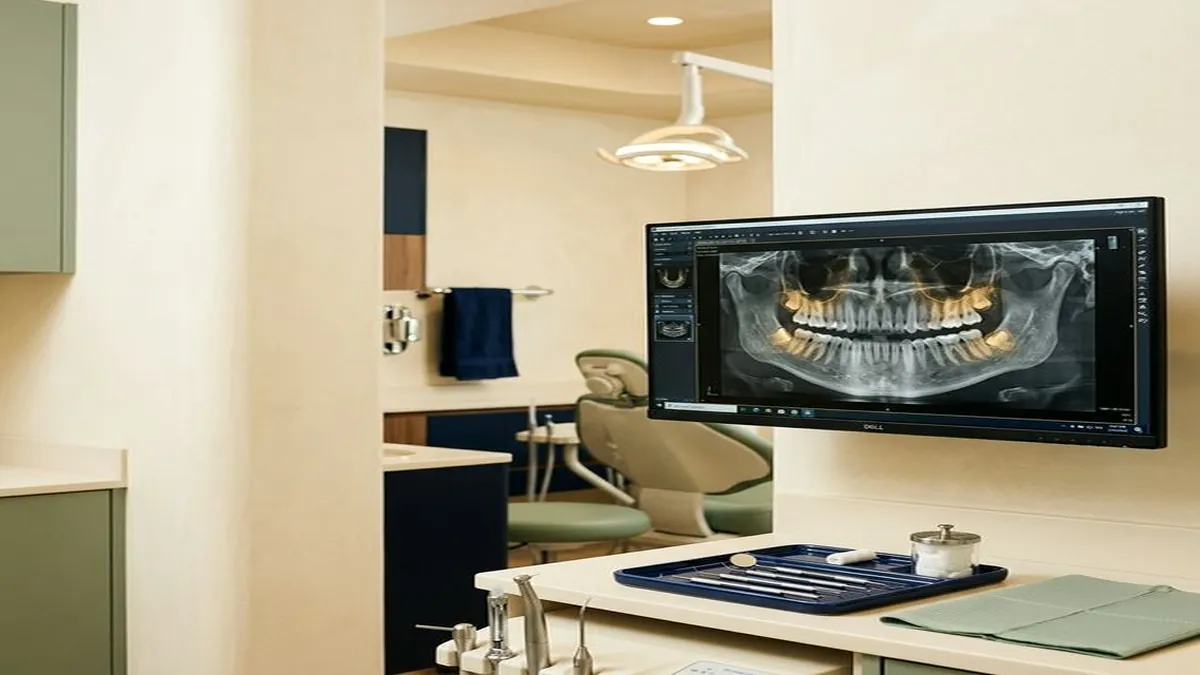
Expect a clinical and radiographic evaluation. Dr. Fatima will probe around the implant to measure pocket depths, assess bleeding, and check for mobility. A periapical X-ray will reveal any bone loss around the implant threads. Together, these findings determine whether the implant is stable, experiencing early peri-implant mucositis, or has progressed to peri-implantitis with bone loss.
Treatment Options for Failing Implants
The treatment approach depends entirely on the type and severity of the problem. Outcomes are significantly better when caught early.
Non-Surgical Debridement
For peri-implant mucositis (inflammation without bone loss), non-surgical debridement, mechanical cleaning of the implant surface and surrounding pockets, is the first-line treatment. Titanium curettes, ultrasonic scalers with specialized implant tips, and air-abrasive devices are used to remove biofilm from the implant surface without scratching it (scratched implant surfaces harbor bacteria more readily). A 2019 systematic review in the Journal of Clinical Periodontology found that non-surgical debridement resolved mucositis in the majority of cases when combined with improved patient hygiene.
Laser Therapy
Laser-assisted decontamination of the implant surface is used as an adjunct to mechanical debridement in peri-implantitis cases. The laser energy kills bacteria within the pockets and decontaminates the implant surface without damaging it. Er:YAG and diode lasers are the most commonly used types for peri-implant treatment.
Antibiotic Therapy
Systemic antibiotics (typically amoxicillin with metronidazole) or locally delivered antibiotics (placed directly into the pocket around the implant) may be prescribed alongside mechanical treatment for active peri-implantitis. Antibiotics alone are not sufficient, they must be combined with mechanical decontamination to be effective.
Surgical Intervention and Bone Grafting
When peri-implantitis has caused significant bone loss, surgical access may be needed to decontaminate the implant surface directly and place bone graft material to regenerate lost bone. The surgical approach involves reflecting the gum tissue, mechanically and chemically cleaning the exposed implant surface, placing bone graft material (often combined with a membrane to contain the graft), and closing the tissue. Results are variable, a 2020 review in Clinical Oral Implants Research found that surgical treatment with bone grafting achieved radiographic bone fill in approximately 50 to 60 percent of treated defects.
Implant Removal (Last Resort)
When an implant is fully mobile, surrounded by extensive bone loss, or associated with a chronic infection that has not responded to treatment, removal is the most responsible option. The implant is reverse-torqued out of the bone (or, if fully loose, simply lifted out), the site is debrided and grafted, and after a healing period of 4 to 6 months, a new implant can often be placed in the regenerated bone.
Implant removal is not a failure of the concept, it is recognition that this particular implant, in this particular situation, is no longer viable. Patients from Shrewsbury and Hopkinton who have had implants placed elsewhere and are experiencing problems often come to Innova Smiles for evaluation and management. Dr. Fatima's FICOI fellowship training includes managing implant complications and re-treatment planning.
How to Prevent Implant Failure
Prevention is far more effective, and far less costly, than treatment. The following habits protect your implant investment for decades:
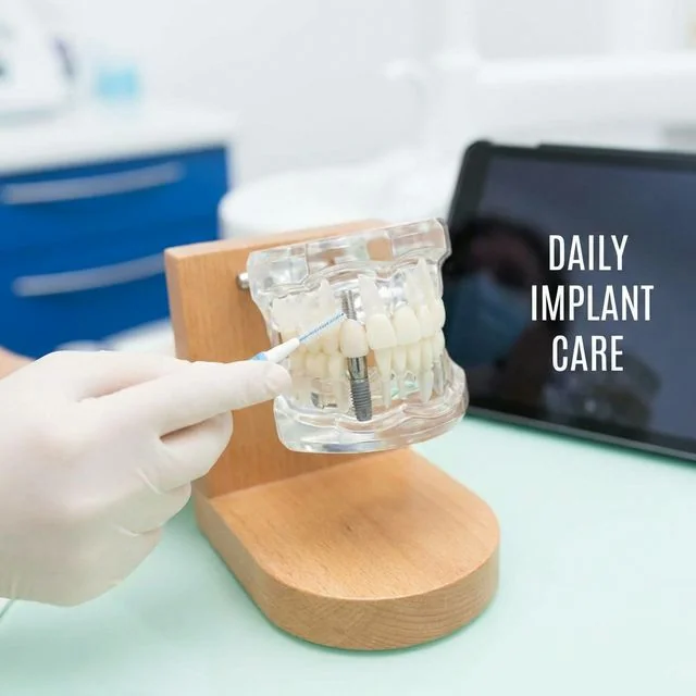
Daily hygiene around implants: Brush twice daily with a soft-bristled toothbrush, paying special attention to the gumline around the implant. Use an interdental brush, floss threader, or water flosser to clean beneath the implant crown where food and bacteria accumulate. Standard dental floss can be used, but implant-specific floss or tape is often easier to maneuver around the abutment.
Regular professional maintenance: The AAID recommends professional cleanings every 3 to 6 months for implant patients. During these visits, your hygienist uses titanium or plastic instruments (not standard steel scalers, which can scratch the implant surface) to clean around the implant and measure pocket depths. Annual radiographs track bone levels over time and detect changes too subtle to feel clinically.
Wear a night guard if you grind: Bruxism is a treatable risk factor. A custom night guard distributes clenching forces and protects both implant crowns and natural teeth from fracture and wear.
Quit smoking: Every cigarette constricts the blood vessels that nourish the bone around your implant. Even after osseointegration is complete, smoking increases peri-implantitis risk throughout the life of the implant.
Manage systemic health conditions: Keep diabetes well controlled, follow medication regimens, and inform your dentist of any changes in your health or medications.
Do not use implant teeth as tools: Avoid biting fishing line, opening packages, cracking nuts, or chewing ice. Implant crowns are strong, but they are not indestructible, and unlike natural teeth, they have no proprioceptive feedback (the nerve signal that tells you how hard you are biting is absent around implants, making it easier to apply excessive force without realizing it).
Success Rate Context
It is worth stepping back from the discussion of failure to put these numbers in perspective. Dental implants remain one of the most successful surgical procedures in medicine. The AAID reports a success rate exceeding 95 percent at 10 years. A 2019 study in Clinical Oral Implants Research tracking over 12,000 implants found a 20-year cumulative survival rate of 93.3 percent. For comparison, hip replacements, widely considered a highly successful procedure, have a 15-year survival rate of approximately 89 percent (per The Lancet, 2019).
When failure does occur, it is usually treatable, often preventable, and rarely a reason to avoid implants as a treatment option. The key is choosing an experienced implant provider, following the post-operative and long-term maintenance protocol, and attending regular follow-up appointments. Understanding the differences between titanium and zirconia implant materials can also help you make informed decisions about your treatment.
Innova Smiles is located at 340 Maple St, Suite 100, Marlborough, MA 01752, and we welcome patients from Sudbury, Natick, and throughout MetroWest Massachusetts for implant consultations, second opinions, and management of implant complications. Call (508) 481-0110 or schedule online.
Concerned about a dental implant? Call (508) 481-0110 or request an evaluation with Dr. Fatima. Early detection makes all the difference.
Related Articles
- Are Dental Implants Permanent? Maintenance Guide
- Dental Implant Recovery: Week-by-Week Timeline
- What to Expect During Dental Implant Surgery