Featured Answer: Why would I need a graft or sinus lift?
After tooth loss, the jawbone can shrink. If there isn’t enough height or width for a stable implant--especially in the upper back jaw near the sinus--a bone graft or sinus lift may be recommended to create a solid foundation and improve long-term success. At Innova Smiles in Marlborough, MA, Dr. Fatima uses CBCT 3D imaging to measure bone dimensions precisely, so MetroWest patients know exactly what to expect before any procedure begins.
Why Bone Loss Happens After Tooth Extraction
Without a tooth root providing mechanical stimulation, the surrounding jawbone begins to resorb through a process called disuse atrophy. Osteoclast cells break down bone tissue faster than osteoblasts can rebuild it, and the imbalance starts within weeks of tooth loss. A landmark study published in Clinical Oral Implants Research (Schropp et al., 2003) documented that patients can lose up to 25% of bone width in the first year alone. Vertical height loss follows in subsequent years, particularly in the posterior maxilla where the sinus cavity expands downward into the vacated space.
The rate of resorption depends on several factors: the location of the missing tooth (upper jaw resorbs faster than lower), how the tooth was lost (traumatic extractions cause more immediate damage), and systemic conditions like osteoporosis or uncontrolled diabetes that impair bone metabolism. A 2017 meta-analysis in the Journal of Dental Research confirmed that alveolar ridge resorption is progressive and irreversible without intervention, with the most dramatic changes occurring in the first six months post-extraction.
For patients in Northborough, Hudson, and surrounding MetroWest communities who have been living with a missing tooth for months or years, bone grafting is often the key to unlocking implant eligibility. The longer you wait, the more bone is lost and the more complex (and costly) the grafting procedure becomes.
What is a Bone Graft for a Dental Implant?
A bone graft is a surgical procedure that adds bone material to your jaw to rebuild the volume lost after extraction or prolonged tooth loss. The graft material serves as an osteoconductive scaffold--a biological framework that encourages your body’s own osteoblast cells to migrate into the area and generate new living bone over several months. This process, called creeping substitution, gradually replaces the graft particles with your own dense, vascularized bone capable of supporting an implant.
The success rate for dental bone grafts is strong. According to a systematic review in the International Journal of Oral & Maxillofacial Implants (Aghaloo & Moy, 2007), grafted sites achieve sufficient bone volume for implant placement in over 90% of cases. Once the graft has matured--typically three to six months after placement--the regenerated bone provides the density, width, and height needed to anchor a titanium implant securely.
Types of Bone Grafts
- Socket preservation (ridge preservation): Performed at the time of extraction to protect the ridge shape. Graft material is placed directly into the empty socket and covered with a collagen membrane, preserving bone width and height for a future implant. This is the most common and least invasive type. A 2019 Cochrane review found that socket preservation reduces horizontal bone loss by approximately 1.5mm compared to unassisted healing--a meaningful difference when every millimeter matters for implant placement.
- Ridge augmentation: Used when the ridge has already narrowed or collapsed after months or years of tooth absence. Additional bone material is placed along the ridge and secured beneath a barrier membrane, increasing width and height to restore the contour needed for both implant stability and natural-looking esthetics. Healing takes four to six months.
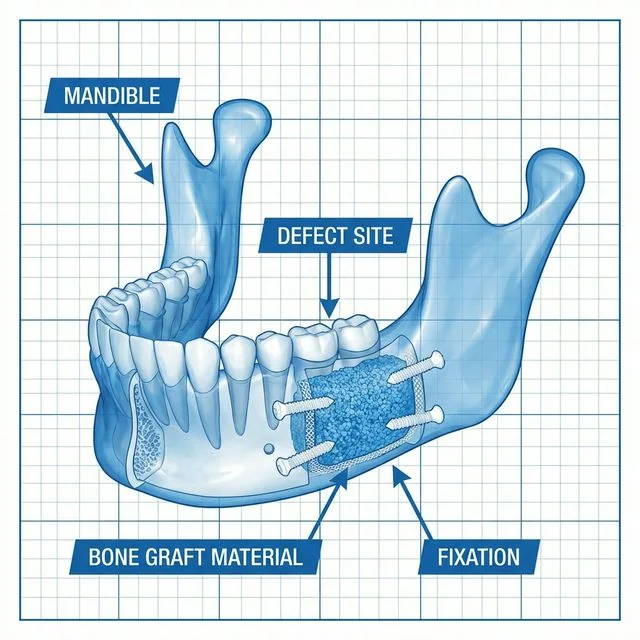
- Block graft: For more significant defects where the ridge has lost substantial three-dimensional volume, a small block of bone (often harvested from the chin, ramus, or back of the jaw) is secured to the deficient site with titanium fixation screws and allowed to integrate over four to six months. Block grafts are particularly useful for patients who need to rebuild both width and height simultaneously.
- Sinus lift (sinus augmentation): Elevates the Schneiderian membrane lining the maxillary sinus in the upper jaw to gain vertical height for upper molar and premolar implants. This is necessary when the sinus floor has pneumatized (dropped) too close to the ridge crest where the implant needs to be placed. It is the most common adjunctive procedure in upper-jaw implant dentistry.
Graft Material Options
Your graft may use one or a combination of these materials, and the choice depends on the defect size, location, and your individual healing profile:
- Autograft (autogenous bone): Bone harvested from your own body--the most-recommended option for osteogenic potential because it contains living cells, growth factors, and a natural scaffold. Common donor sites include the chin symphysis, mandibular ramus, and for larger grafts, the iliac crest (hip). The downside is a second surgical site, which adds discomfort and healing time.
- Allograft (donor bone): Processed human bone from an accredited tissue bank. The American Academy of Implant Dentistry (AAID) notes that allografts are the most widely used graft material in the United States and are rigorously screened and sterilized. They provide an excellent osteoconductive scaffold without requiring a donor-site surgery. Demineralized bone matrix (DBM) is a common allograft form that retains bone morphogenetic proteins (BMPs) to stimulate new bone formation.
- Xenograft: Bone-derived mineral from animal sources (typically bovine, such as Bio-Oss), processed to remove all organic components while preserving the natural hydroxyapatite structure. Xenografts resorb slowly, which makes them excellent space-maintainers for sinus lifts and large ridge augmentations. A 10-year follow-up study in Clinical Oral Implants Research (Jensen et al., 2012) demonstrated stable bone levels around implants placed in xenograft-augmented sinuses.
- Alloplast (synthetic bone): Laboratory-manufactured materials such as beta-tricalcium phosphate (beta-TCP) or calcium phosphate ceramics that mimic natural bone mineral. These are completely biocompatible and eliminate any concern about disease transmission. They are commonly used in socket preservation and smaller defects.
Dr. Fatima selects the material best suited to each patient’s clinical situation. In many cases, a combination approach--for example, allograft mixed with platelet-rich fibrin (PRF) from your own blood--produces faster, denser bone regeneration.
When is a Sinus Lift Needed?
A sinus lift is specifically indicated for the upper back jaw (premolars and molars), where the maxillary sinuses sit directly above the tooth roots. When a posterior tooth is lost, the sinus floor gradually drops into the vacated space through a process called sinus pneumatization. Over time, this leaves too little bone height between the ridge crest and the sinus membrane to safely place an implant.
If CBCT imaging shows less than 8 to 10 millimeters of bone height between the ridge crest and the sinus floor, a sinus lift is typically recommended before or during implant placement. There are two approaches:
Lateral Window Sinus Lift
This is the traditional technique for significant height deficiency (less than 5mm of residual bone). The surgeon creates a small window in the lateral wall of the sinus, carefully elevates the Schneiderian membrane, and packs graft material beneath it. Healing takes five to eight months before implant placement. According to a systematic review in the Journal of Clinical Periodontology (Pjetursson et al., 2008), implants placed with lateral window sinus lift procedures achieved a 3-year survival rate of 90.1% (95% CI: 86.4%-92.8%), with the best outcomes (98.3%) achieved using rough-surface implants and membrane coverage of the lateral window.
Transcrestal (Osteotome) Sinus Lift
When residual bone height measures 5 to 8mm, a less invasive crestal approach may be used. The sinus floor is gently elevated through the implant preparation site using specialized osteotome instruments, and graft material is placed through the same opening. In many cases, the implant can be placed simultaneously, eliminating a separate grafting surgery.
For MetroWest patients traveling from Sudbury or Southborough, the crestal technique can mean fewer office visits and a shorter overall treatment timeline. Dr. Fatima evaluates which approach is appropriate based on your 3D imaging.
Bone Grafting Cost: What to Expect in Massachusetts
Bone grafting costs vary based on the type and complexity of the procedure. Here is what patients in the Marlborough and MetroWest area can typically expect:
Most PPO dental insurance plans cover a portion of bone grafting when it is medically necessary for implant placement. Coverage typically falls under major restorative benefits, and we submit pre-authorization to your carrier before scheduling surgery. We also offer 0% financing through CareCredit and Cherry for patients who want to spread the cost over monthly payments. Visit our insurance and financing page for details.
What to Expect During and After the Procedure
- According to the American Association of Oral and Maxillofacial Surgeons (AAOMS), local anesthesia with comfort-focused protocols is the standard approach; comfort options including premium topical numbing and gentle local anesthesia are available for patients who prefer extra support
- The procedure itself typically takes 45 to 90 minutes depending on the type and extent of grafting
- Mild to moderate swelling and discomfort for three to five days, managed with prescribed or over-the-counter medications. Swelling peaks around day two to three and then gradually subsides
- Nasal precautions after a sinus lift: avoid blowing your nose forcefully, sneeze with your mouth open, and skip straw use for two weeks to protect the elevated membrane
- Healing time of three to six months before the implant can be placed, though simultaneous placement is sometimes possible for socket preservation grafts and transcrestal sinus lifts
- Follow-up imaging (periapical or CBCT) at three to four months to confirm bone maturation and readiness for the implant phase
- Antibiotics are prescribed prophylactically for sinus lifts and larger grafts; a chlorhexidine rinse is typically recommended for the first two weeks
Your fellowship advantage: Dr. Fatima’s FICOI fellowship training supports precise diagnostics and minimally invasive techniques to reduce surgical trauma and enhance predictability. Fellowship-trained implant dentists have completed hundreds of supervised grafting cases beyond standard dental school training.
Technology That Makes a Difference
At Innova Smiles, we rely on CBCT (cone beam computed tomography) to create a three-dimensional map of your jaw before any grafting procedure. Unlike traditional two-dimensional X-rays, CBCT imaging provides sub-millimeter measurements of bone height, width, and density at every potential implant site. This allows Dr. Fatima to identify the exact areas of bone deficiency, measure distances to critical structures like the inferior alveolar nerve and the sinus floor, and plan the most conservative approach possible.
We also use digital surgical guides fabricated from CBCT data to execute the planned implant positions with precision. A systematic review in Clinical Oral Implants Research (Tahmaseb et al.) found that guided surgery achieved a mean accuracy of 1.2mm at the implant tip--a significant improvement over freehand placement that directly reduces the risk of complications.
If you are also exploring what to expect during implant surgery, this same imaging guides every step of the process.
How to Know If You Need a Bone Graft
Not every implant patient requires bone grafting. Approximately 50% of single-tooth implant cases can proceed without any grafting at all, provided the tooth was extracted atraumatically and the site has not been edentulous for an extended period. The need is determined by your CBCT scan results, specifically the measurements of bone height, width, and density at the planned implant site. If your bone dimensions meet the minimum thresholds--typically at least 10mm of height and 6mm of width--a graft may not be necessary.
Factors that increase the likelihood of needing a bone graft for a dental implant include:
- Teeth that have been missing for more than a year, allowing significant resorption
- A history of periodontal disease causing bone destruction around neighboring teeth
- Previous denture wear, which accelerates ridge resorption through constant pressure
- Traumatic tooth loss or surgical extraction that damaged the buccal plate
- Naturally thin bone anatomy in the upper jaw, particularly the premolar and molar regions
- Systemic conditions such as osteoporosis that compromise bone density
- Smoking, which impairs blood flow and slows bone healing by 30 to 40% according to research in The Journal of the American Dental Association
Dr. Fatima reviews your 3D imaging with you at your consultation, pointing out exactly where bone is sufficient and where augmentation may be needed. This transparent approach ensures you understand every step of the treatment plan before committing to surgery.
Healing Timeline: What Happens After a Bone Graft
Understanding the biological timeline helps set realistic expectations:
During the healing period, the graft site must remain undisturbed. Removable prosthetics (flippers or partial dentures) are adjusted to avoid pressure on the graft, and hard or crunchy foods should be avoided on that side. Most patients report that the recovery is milder than they expected--comparable to a routine extraction.
Your Questions, Answered (Marlborough and MetroWest)
We will review 3D imaging, your timeline, and budget during a thorough consultation. Many patients along the I-495 corridor--from Framingham to Westborough--prefer sequencing that minimizes time off work. We coordinate grafting and implant phases to fit your schedule and maximize insurance coverage when applicable. Patients from Shrewsbury, Grafton, and Hopkinton also visit our Marlborough office for implant consultations because many grafting and implant phases can be coordinated here; when a case benefits from additional expertise, we coordinate referral or co-management with an appropriate specialist.
Do bone grafts hurt?
Most patients report less discomfort than they anticipated. Local anesthesia keeps you comfortable during the procedure, and post-operative pain is typically managed with ibuprofen and acetaminophen on an alternating schedule. Sinus lifts tend to produce more swelling than socket grafts, but pain levels are usually moderate and well-controlled.
Can I get a bone graft and implant at the same time?
In some cases, yes. When residual bone provides enough initial stability (at least 4 to 5mm of height and width), Dr. Fatima can place the implant and pack graft material around it simultaneously. This combined approach reduces the total number of surgeries and shortens the overall treatment timeline by several months.
What is the success rate of bone grafts for dental implants?
Published literature reports bone graft success rates between 90% and 97%, depending on the graft type, patient health, and surgical technique. Smokers and patients with uncontrolled diabetes have lower success rates, which is why we address these risk factors during your treatment planning.
How long do I have to wait between graft and implant?
Typically three to six months, verified by follow-up imaging. Socket preservation grafts often heal faster (three to four months), while sinus lifts and block grafts may need five to eight months of maturation.
Considering implants but unsure about bone grafting? Call (508) 481-0110 or request a consultation. We provide complimentary CBCT imaging with every implant consultation so you get a clear picture of your bone health from the first visit.
Related Articles
- Am I a Candidate for Dental Implants?
- Guided Implant Surgery: CBCT Precision and Safety
- Dental Implant Recovery: Day-by-Day Timeline
Related Services
Sources & Further Reading
- The accuracy of static computer-aided implant surgery: A systematic review and meta-analysis — PubMed/Clinical Oral Implants Research