
Featured Answer: Are dental X-rays safe?
Yes. Digital X-rays use a very low dose of radiation, far less than older film systems and well within safety guidelines established by the ADA and the FDA. At Innova Smiles in Marlborough, MA, Dr. Fatima uses the latest digital imaging technology to find problems that are not visible during a visual exam while keeping radiation exposure as low as reasonably achievable. Patients from Shrewsbury, Northborough, and across MetroWest can feel confident that their diagnostic imaging is both safe and essential.
Why X-Rays Matter
Many dental problems develop silently beneath the surface. Without X-rays, cavities between teeth, infections at the root tip, bone loss from periodontal disease, and developing wisdom teeth can go undetected until they cause pain or require emergency treatment. A 2012 study in Dentomaxillofacial Radiology found that bitewing radiographs detected 40 to 60 percent more interproximal carious lesions than clinical examination alone, meaning dentists who skip imaging miss roughly half the cavities hiding between teeth. Early detection through routine imaging saves time, money, and discomfort. As we discuss in our post on the link between oral health and overall wellness, catching problems early has benefits that extend well beyond the mouth.
Consider a common scenario we see in our Marlborough office: a patient comes in with no visible issues, no pain, and good brushing habits. A set of bitewings reveals a small cavity forming between two molars, invisible to the naked eye. Caught at this stage, a simple filling resolves the problem in under 30 minutes. Left undetected for another year, that same cavity penetrates into the nerve chamber, requiring a root canal, a crown, and several additional appointments. Dental X-ray radiation from those diagnostic bitewings is trivially small compared to the cost and discomfort of advanced treatment.
Types of Dental X-Rays
Different clinical situations call for different types of images. Understanding what each type shows helps you appreciate why Dr. Fatima may recommend one over another:
- Bitewings: The most common type taken during routine checkups. Bitewings show the upper and lower back teeth in a single image and are ideal for detecting cavities between teeth and monitoring bone levels. A standard set of four bitewings covers both sides of the mouth and takes less than 60 seconds to capture with digital sensors.
- Periapical: These capture the entire tooth from crown to root tip and the surrounding bone. They are essential for diagnosing infections, abscesses, and root fractures. After a root canal or implant placement, periapical films help us verify healing and integration.
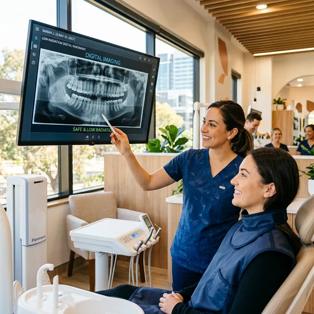
- Panoramic (Panorex): A single image that shows all teeth, both jaws, the sinuses, and the temporomandibular joints. Panoramic X-rays are useful for orthodontic planning, implant evaluation, and wisdom tooth assessment. The camera rotates around your head while you stand still, and the entire process takes about 20 seconds.
- CBCT (Cone Beam CT): A three-dimensional scan that provides detailed views of bone density, nerve pathways, and tooth anatomy. Dr. Fatima uses CBCT imaging for precise implant placement and complex treatment planning. A single CBCT scan delivers roughly 0.05 to 0.5 mSv depending on the field of view, which is still a fraction of a medical CT scan of the head (approximately 2.0 mSv).
- Occlusal: A larger film placed on the biting surface captures the full arch. Occlusal views are helpful for locating impacted teeth, detecting jaw fractures, or evaluating palatal pathology in children.
How Often Should You Get Dental X-Rays?
The frequency depends on your individual risk factors. The ADA and the FDA jointly published updated selection criteria in 2012 that remain the standard of care. Those guidelines recommend imaging intervals based on clinical risk rather than arbitrary timelines:
- Low risk (healthy adults with no active decay or gum disease): Bitewings every 24 to 36 months
- Moderate risk (history of cavities or early gum disease): Bitewings every 12 to 18 months
- High risk (active decay, periodontal disease, or complex treatment): Bitewings every 6 to 12 months
- New patients: A full-mouth series or panoramic image at the initial visit to establish a baseline
- Children: Frequency is based on age, cavity risk, and eruption patterns. The American Academy of Pediatric Dentistry recommends bitewings as early as the eruption of adjacent primary molars if the child is at elevated caries risk
Dr. Fatima follows these evidence-based guidelines and only prescribes X-rays when the diagnostic benefit outweighs the minimal exposure. Every image we take answers a specific clinical question.
Radiation: Putting the Numbers in Perspective
Understanding how dental X-ray radiation compares to everyday exposure helps put safety concerns in context:
| Source | Approximate Dose |
|---|---|
| Single digital dental X-ray | 0.005 mSv |
| Set of 4 bitewings | 0.005 mSv |
| Daily natural background radiation | 0.008 mSv |
| Cross-country flight (NY to LA) | 0.04 mSv |
| Panoramic dental X-ray | 0.01–0.03 mSv |
| CBCT dental scan (small field) | 0.05–0.5 mSv |
| Chest X-ray | 0.10 mSv |
| Annual background radiation (U.S. average) | 3.0 mSv |
| Medical CT of the head | 2.0 mSv |
A set of four digital bitewings delivers roughly the same radiation as a couple of days of natural background exposure or half a cross-country flight. According to the American Dental Association (ADA), digital sensors require up to 80 percent less radiation than traditional film, making modern dental imaging one of the lowest-dose diagnostic tools in medicine. The National Council on Radiation Protection and Measurements (NCRP Report No. 177, 2019) confirmed that dental imaging contributes less than 2.5 percent of the total medical radiation exposure received by the U.S. population.
To put this in personal terms: if you live in the MetroWest area and fly out of Boston Logan once or twice a year for a family vacation, the cosmic radiation from those flights exceeds your annual dental X-ray dose.
Digital vs. Film: Why Digital Wins
The transition from film to digital imaging represents one of the most significant safety and quality improvements in modern dentistry. Here is how the two technologies compare:
- Up to 80% less radiation: Digital sensors are far more sensitive than film, requiring a smaller dose to produce a clear image. A study published in Oral Surgery, Oral Medicine, Oral Pathology, Oral Radiology and Endodontology confirmed that digital receptors consistently required lower exposure settings than D-speed and E-speed film.
- Instant results: Images appear on the screen in seconds, eliminating chemical processing and wait times. This means Dr. Fatima can review findings with you in real time, pointing out areas of concern on the chairside monitor.
- Enhanced detail: Software allows us to adjust contrast, zoom in on areas of concern, and highlight subtle changes. Color mapping and density measurement tools let us quantify bone loss or track lesion size between visits.
- Fewer retakes: Immediate review means we can confirm image quality before you leave the chair. With film, an underexposed or poorly positioned image was not discovered until after chemical development, requiring a second exposure.
- Secure sharing: Digital files are transmitted instantly to specialists, insurance companies, or your new dentist if you relocate. Patients who move within MetroWest, from Framingham to Westborough, can have their records transferred electronically the same day.
- Eco-friendly: No chemical developing solutions, lead foil, or plastic film waste. Traditional film processing used fixer and developer chemicals classified as hazardous waste.
- Long-term archiving: Digital images are stored indefinitely without degradation. Film deteriorates over time and requires physical storage space.
How Digital Dental X-Rays Work
A digital X-ray sensor is a small, flat electronic plate that replaces the film packet. When X-ray photons strike the sensor, they generate an electronic signal that is converted into a grayscale image by the software. The entire exposure lasts a fraction of a second. Phosphor plate systems, an alternative digital technology, use a flexible plate that is scanned by a laser reader after exposure. Both methods produce high-resolution diagnostic images at radiation doses well below those required for conventional film.
At Innova Smiles, we use direct digital sensors for intraoral images because they offer the fastest image acquisition and the lowest patient dose. For panoramic imaging, our unit uses a digital receptor with automatic exposure optimization that adjusts the dose based on patient size.
Dental X-Rays During Pregnancy
Both the ADA and the American College of Obstetricians and Gynecologists (ACOG) agree that dental X-rays with proper shielding are safe during pregnancy. The FDA further confirms that modern digital X-ray doses are well within safe limits for expectant mothers. A lead apron and thyroid collar are always used to protect the patient.
A 2013 position statement from ACOG specifically noted that diagnostic dental radiography during pregnancy is safe and should not be withheld when clinically indicated. The radiation dose from a full set of dental X-rays (approximately 0.1 mSv) is far below the 50 mSv threshold at which fetal effects have been observed in animal studies.
However, elective or routine imaging is typically postponed until after delivery when possible. If you are pregnant or think you may be, let our team know so we can tailor your visit accordingly. Urgent diagnostic imaging should never be delayed, as untreated dental infections can pose a greater risk to both mother and baby than the minimal radiation involved. Periodontal infections during pregnancy have been associated with preterm birth and low birth weight in studies published in the Journal of Periodontology.
Dental X-Rays for Children: What Parents Need to Know
Parents in our Marlborough office frequently ask whether dental X-rays are safe for their children. The answer is yes, with appropriate precautions. Children actually benefit significantly from imaging because their teeth and jaws are still developing, and early detection of problems like ectopic tooth eruption, supernumerary teeth, or early cavities can prevent more invasive treatment later.
Key considerations for pediatric dental imaging:
- Dose adjustment: Our digital X-ray equipment automatically reduces the exposure settings for smaller patients. A child receives a lower dose per image than an adult.
- Thyroid protection: The thyroid gland is more radiosensitive in children. We use a thyroid collar for every pediatric exposure, consistent with ADA recommendations updated in 2012.
- Frequency: The ADA recommends that children at high caries risk (visible plaque, frequent snacking, limited fluoride exposure) receive bitewings every 6 to 12 months. Low-risk children may only need bitewings every 12 to 24 months.
- First X-rays: Most children receive their first bitewing images around age 5 or 6, when the primary molars are in contact and interproximal cavities become a risk. A panoramic image around age 6 to 7 evaluates the developing permanent dentition.
Many parents wonder how to prepare their child for dental X-rays. We recommend telling children that the X-ray camera will take a quick picture of their teeth to help Dr. Fatima make sure everything is growing correctly. The sensor is small, the exposure lasts less than a second, and there is no pain involved. For younger children who have difficulty holding the sensor, we may use a panoramic image instead, which requires no intraoral sensor at all.
Local Perspective
Busy families in Marlborough, Hudson, and Southborough value the speed and clarity of digital imaging during routine checkups. If you are a working parent fitting a cleaning into a lunch break off Route 20, or a retiree who wants thorough preventive care, you will get low-dose, high-accuracy diagnostics at every visit.
MetroWest residents also benefit from our investment in CBCT technology. Rather than referring patients to a hospital radiology center for 3D imaging--which can mean driving to Worcester or Boston, waiting weeks for an appointment, and paying a separate facility fee--we capture CBCT scans in our Marlborough office during the same visit. This saves time, reduces cost, and eliminates the hassle of coordinating between providers.
ALARA Principle: As Low As Reasonably Achievable
At Innova Smiles, we follow the ALARA principle endorsed by both the ADA and the National Council on Radiation Protection (NCRP). This means we use every available technique to minimize radiation exposure while still obtaining diagnostically useful images. Specific measures include:
- Rectangular collimation, narrows the X-ray beam to the size of the sensor, reducing the area of tissue exposed by up to 60% compared to round collimation. A 2009 study in the Journal of Dental Research confirmed that rectangular collimation reduced patient dose by up to five times compared to round collimation with no loss of diagnostic quality.
- Lead aprons and thyroid collars, standard for all patients during any imaging procedure
- Digital sensors, require significantly lower radiation doses than traditional film
- Selection criteria: X-rays are prescribed based on individual clinical need, not on a rigid calendar schedule
- Proper technique, accurate beam alignment and sensor positioning minimize the need for retakes
- Equipment maintenance, our X-ray units are inspected and calibrated annually per Massachusetts Department of Public Health regulations to ensure they deliver consistent, accurate doses
Common Questions About Dental X-Ray Safety
Do dental X-rays cause cancer? The risk from dental X-rays is extremely low. A 2012 study in Cancer that received significant media attention suggested a link between frequent dental X-rays and meningioma, but subsequent analysis by the ADA noted critical methodological limitations, including reliance on patient recall of imaging history over decades. The consensus among radiation safety organizations, including the Health Physics Society, is that risks below 50 mSv per year (2,500 times the dose of a full set of dental X-rays) are too small to be observed and may be nonexistent.
Can I refuse dental X-rays? You always have the right to decline any diagnostic procedure. However, without current imaging, Dr. Fatima cannot diagnose conditions that are invisible to the naked eye. Declining X-rays may mean that cavities, infections, or bone loss progress undetected until they cause pain or require more extensive treatment. We encourage patients to discuss their concerns so we can explain exactly why a specific image is recommended.
Are dental X-rays safe for patients with thyroid conditions? Yes. The dose to the thyroid from a dental X-ray is negligible, and we use thyroid collars as standard practice. Patients with thyroid disease, including those who have undergone radioactive iodine treatment, can safely receive dental imaging. If you have concerns, bring them up at your appointment and we will walk through the specifics.
What about cumulative radiation exposure? The concept of cumulative dose is often misunderstood. While it is true that radiation effects are cumulative over a lifetime, the doses from dental imaging are so small that they represent a negligible fraction of total lifetime exposure. The average American receives about 6.2 mSv per year from all sources combined (natural background, medical imaging, consumer products). A full year of dental X-rays adds less than 0.1 mSv to that total.
The Future of Dental Imaging
Digital imaging continues to advance. Emerging technologies like artificial intelligence-assisted image analysis are showing promise in detecting cavities and bone loss with accuracy that matches or exceeds trained clinicians. A 2020 systematic review in Journal of Dental Research found that deep-learning algorithms achieved sensitivity above 90 percent for caries detection on bitewing radiographs. At Innova Smiles, we stay current with evidence-based imaging technology so MetroWest patients benefit from the safest, most accurate diagnostics available.
Photon-counting detector technology, currently in development for dental applications, promises to further reduce doses while improving image contrast. As these tools become available, we will evaluate them against the same ALARA standards we apply to every imaging decision today.
Need updated X-rays or have questions about dental X-ray radiation safety? Call (508) 481-0110 or book now. Our Marlborough team is always happy to walk you through exactly what we are looking for and why.
Related Articles
- Why Dental Cleanings Matter More Than You Think
- Why Oral Cancer Screenings Are Essential
- Digital Dental Impressions: Gag-Free Comfort